Elbow Sprain in a Youngster
Radiology Cases in Pediatric Emergency Medicine
Volume 1, Case 17
Loren G. Yamamoto, MD, MPH
Kapiolani Medical Center For Women And Children
University of Hawaii John A. Burns School of Medicine
An 8 year old female presents to the ED after falling
off the monkey bars at school, complaining of pain in
her elbow.
Exam: There is tenderness over the lateral aspect
of the elbow and reduced range of motion on flexion,
supination, and pronation due to pain. Distally, the
exam is negative. Possibly, there is some very mild
elbow swelling, but this is not obvious. There are no
deformities. Sensation is intact distally. The radial
pulse is present at the wrist. The proximal humerus,
shoulder, and clavicles are non-tender. Radiographs of
the elbow are obtained.
View elbow radiographs.
 These radiographs show a very subtle fracture of
the radial head at the metaphysis. This can be seen as
a slight interruption in the smooth contour of the
metaphysis. The anterior fat pad is prominent. Click on
[Enlarge] to magnify the image. The radial head
fracture is best seen on the AP view on the lateral size
(left on the screen) of the radial metaphysis as a slight
irregularity in the cortex.
An additional oblique view shows the fracture
somewhat better.
View oblique view.
These radiographs show a very subtle fracture of
the radial head at the metaphysis. This can be seen as
a slight interruption in the smooth contour of the
metaphysis. The anterior fat pad is prominent. Click on
[Enlarge] to magnify the image. The radial head
fracture is best seen on the AP view on the lateral size
(left on the screen) of the radial metaphysis as a slight
irregularity in the cortex.
An additional oblique view shows the fracture
somewhat better.
View oblique view.
 Discussion & Teaching Points:
1) A radial head fracture is not always easy to
appreciate. The patient's symptoms may be relatively
mild. Flexion and extension of the elbow are frequently
unremarkable. Supination and pronation may be more
limited and/or painful. There is often tenderness over
the radial head. Since radial head fractures tend to
occur in older individuals, it is unlikely to be mistaken
for a subluxed radial head as in a nursemaid's elbow;
however, since it can occur in younger children also,
this diagnosis should still be considered if the history
does not suggest a classic pulling injury.
2) A sprain injury of the elbow is a diagnostic pitfall
that should be avoided. This patient does NOT have an
elbow sprain. Occult fractures in the elbow are often
present and difficult to appreciate radiographically. A
normal set of radiographs, even after review by
radiologists, are not able to totally rule out a fracture. It
may be useful to routinely issue a standardized
instruction sheet explaining this possibility to patients.
Refer to Case 19 (Swollen Elbow With a Normal X-ray).
3) Elbow injuries are fracture prone. Radiographs
of this area can be difficult to interpret unless a
methodical review of the radiographs is performed.
One of the things that should be carefully inspected is
the contour of the radial head. The slope from the
diaphysis to the metaphysis towards the radial head
should be gradual and smooth with no sharp angles
associated with it. It should NOT look like the end of a
baseball bat (handle end).
View radiograph in a teenager.
Discussion & Teaching Points:
1) A radial head fracture is not always easy to
appreciate. The patient's symptoms may be relatively
mild. Flexion and extension of the elbow are frequently
unremarkable. Supination and pronation may be more
limited and/or painful. There is often tenderness over
the radial head. Since radial head fractures tend to
occur in older individuals, it is unlikely to be mistaken
for a subluxed radial head as in a nursemaid's elbow;
however, since it can occur in younger children also,
this diagnosis should still be considered if the history
does not suggest a classic pulling injury.
2) A sprain injury of the elbow is a diagnostic pitfall
that should be avoided. This patient does NOT have an
elbow sprain. Occult fractures in the elbow are often
present and difficult to appreciate radiographically. A
normal set of radiographs, even after review by
radiologists, are not able to totally rule out a fracture. It
may be useful to routinely issue a standardized
instruction sheet explaining this possibility to patients.
Refer to Case 19 (Swollen Elbow With a Normal X-ray).
3) Elbow injuries are fracture prone. Radiographs
of this area can be difficult to interpret unless a
methodical review of the radiographs is performed.
One of the things that should be carefully inspected is
the contour of the radial head. The slope from the
diaphysis to the metaphysis towards the radial head
should be gradual and smooth with no sharp angles
associated with it. It should NOT look like the end of a
baseball bat (handle end).
View radiograph in a teenager.
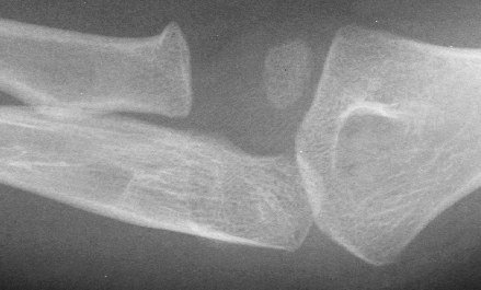
 This radiograph shows a subtle fracture of the radial
head. Note the very slight corner interrupting the
smooth contour of the radial head metaphysis. The
radial head appears to resemble a knob on a pole
rather than a funnel shape. The smooth contour of the
radial head as one proceeds from the epiphysis to the
metaphysis should be carefully inspected. Any angles
noted in this smooth progression may represent a
fracture, especially if the clinical findings confirm this.
Enlarging the radiograph or examining the
radiograph with a magnifying glass may make it
easier to identify these.
View radiograph in a toddler.
This radiograph shows a subtle fracture of the radial
head. Note the very slight corner interrupting the
smooth contour of the radial head metaphysis. The
radial head appears to resemble a knob on a pole
rather than a funnel shape. The smooth contour of the
radial head as one proceeds from the epiphysis to the
metaphysis should be carefully inspected. Any angles
noted in this smooth progression may represent a
fracture, especially if the clinical findings confirm this.
Enlarging the radiograph or examining the
radiograph with a magnifying glass may make it
easier to identify these.
View radiograph in a toddler.
 This radiograph shows a fracture of the radial head.
Note the sharp angle seen at the radial head
metaphysis. This patient presented with symptoms
similar to a nursemaid's elbow; however, the history did
not suggest a pulling injury.
This radiograph shows a fracture of the radial head.
Note the sharp angle seen at the radial head
metaphysis. This patient presented with symptoms
similar to a nursemaid's elbow; however, the history did
not suggest a pulling injury.
Return to Radiology Cases In Ped Emerg Med Case Selection Page
Return to Univ. Hawaii Dept. Pediatrics Home Page