Test Your Skill In Reading Pediatric Lateral Necks
Radiology Cases in Pediatric Emergency Medicine
Volume 2, Case 20
Loren G. Yamamoto, MD, MPH
Kapiolani Medical Center For Women And Children
University of Hawaii John A. Burns School of Medicine
Interpreting lateral neck films usually involves
children with respiratory infections, foreign bodies, or
cervical spine conditions. Case 10 in Volume 1
reviewed the radiographic findings distinguishing
several types of respiratory infections that result in
airway symptoms (croup, retropharyngeal abscess, and
epiglottitis). Case 8 in Volume 1 reviewed some of the
clinical and radiographic features of airway foreign
bodies. Cases 1 and 7 in Volume 2 reviewed some of
the complications of esophageal and bronchial foreign
bodies. Case 5 in Volume 1 reviewed the radiographic
features of C2-C3 pseudosubluxation versus true
subluxation. With this background information, 16
lateral neck radiographs are contained in this case for
review to test your interpretation skills. No clinical
information is given here. Some of these films are soft
tissue studies, while others are cervical spine studies.
To conserve disk space, the images are limited to the
area of interest only.
A general approach to reviewing these radiographs
can be more consistent if one adheres to a standard
method of review.
1. Bony Alignment: Line up the anterior borders of
the vertebral bodies, the posterior borders of the
vertebral bodies, the vertebral arches, and the spinous
processes.
2. Height of the vertebral bodies and disk spaces.
3. Relationship of the odontoid (C2) and the atlas
(C1).
4. Positioning of the neck: Is the neck in flexion,
extension, or neutral.
5. Width of the prevertebral soft tissue space. This
thickness is usually half the width of a vertebral body
and should not exceed the width of a vertebral body.
6. Epiglottis: Examine the shape of the epiglottis
and the size of the pre-epiglottic space (vallecula).
7. Subglottic airway size.
View Case A.
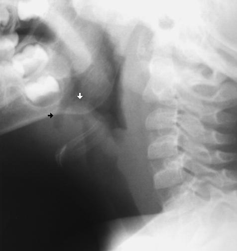 Interpretation of Case A
1. Bony Alignment: Normal. Not able to see C7.
2. Vertebral bodies and disk spaces: Normal sizes.
Not able to see C7.
3. C1-C2: Normal.
4. Positioning: Extension.
5. Prevertebral space: Slightly full, but less than the
width of a vertebral body.
6. Epiglottis: Thumb-like in appearance (white
arrow). It should normally appear thin or triangular.
The pre-epiglottic space (black arrow) is narrow and
nearly obliterated.
7. Subglottic airway size: Satisfactory.
Impression: Epiglottitis.
View Case B.
Interpretation of Case A
1. Bony Alignment: Normal. Not able to see C7.
2. Vertebral bodies and disk spaces: Normal sizes.
Not able to see C7.
3. C1-C2: Normal.
4. Positioning: Extension.
5. Prevertebral space: Slightly full, but less than the
width of a vertebral body.
6. Epiglottis: Thumb-like in appearance (white
arrow). It should normally appear thin or triangular.
The pre-epiglottic space (black arrow) is narrow and
nearly obliterated.
7. Subglottic airway size: Satisfactory.
Impression: Epiglottitis.
View Case B.
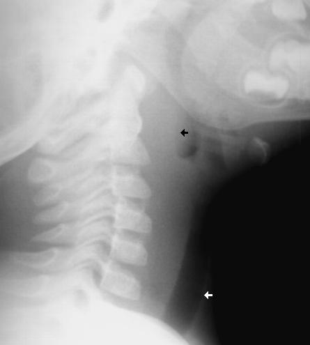 Interpretation of Case B
1. Bony Alignment: Normal. Not able to see C7.
2. Vertebral bodies and disk spaces: Normal sizes.
Not able to see C7.
3. C1-C2: Normal.
4. Positioning: Extension.
5. Prevertebral space: Widened (black arrow). It is
slightly thicker than the width of a vertebral body.
6. Epiglottis: Thin. The pre-epiglottic space is
normal (wider than in Case A).
7. Subglottic airway size: Satisfactory (white
arrow).
Impression: Retropharyngeal abscess.
View Case C.
Interpretation of Case B
1. Bony Alignment: Normal. Not able to see C7.
2. Vertebral bodies and disk spaces: Normal sizes.
Not able to see C7.
3. C1-C2: Normal.
4. Positioning: Extension.
5. Prevertebral space: Widened (black arrow). It is
slightly thicker than the width of a vertebral body.
6. Epiglottis: Thin. The pre-epiglottic space is
normal (wider than in Case A).
7. Subglottic airway size: Satisfactory (white
arrow).
Impression: Retropharyngeal abscess.
View Case C.
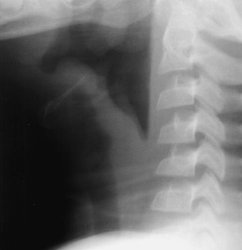
 Interpretation of Case C
1. Bony Alignment: Normal. Not able to see C7.
Spinous processes are not included in the image.
2. Vertebral bodies and disk spaces: Normal sizes.
Not able to see C7.
3. C1-C2: Normal.
4. Positioning: Extension.
5. Prevertebral space: Not widened.
6. Epiglottis: Thin. The pre-epiglottic space is
normal.
7. Subglottic airway size: Narrowed.
Impression: Subglottic edema. Croup.
View Case D.
Interpretation of Case C
1. Bony Alignment: Normal. Not able to see C7.
Spinous processes are not included in the image.
2. Vertebral bodies and disk spaces: Normal sizes.
Not able to see C7.
3. C1-C2: Normal.
4. Positioning: Extension.
5. Prevertebral space: Not widened.
6. Epiglottis: Thin. The pre-epiglottic space is
normal.
7. Subglottic airway size: Narrowed.
Impression: Subglottic edema. Croup.
View Case D.
 Interpretation of Case D
1. Bony Alignment: Normal.
2. Vertebral bodies and disk spaces: Normal sizes.
Not able to fully see C7.
3. C1-C2: Probably normal, difficult to see.
4. Positioning: Extension.
5. Prevertebral space: Borderline widening. It is
slightly less wide than the width of a vertebral body. It
is clearly wider than half of a vertebral body.
6. Epiglottis: Thin. The pre-epiglottic space is
normal.
7. Subglottic airway size: Image is too dark to see
this.
Impression: Possible early retropharyngeal abscess.
View Case E.
Interpretation of Case D
1. Bony Alignment: Normal.
2. Vertebral bodies and disk spaces: Normal sizes.
Not able to fully see C7.
3. C1-C2: Probably normal, difficult to see.
4. Positioning: Extension.
5. Prevertebral space: Borderline widening. It is
slightly less wide than the width of a vertebral body. It
is clearly wider than half of a vertebral body.
6. Epiglottis: Thin. The pre-epiglottic space is
normal.
7. Subglottic airway size: Image is too dark to see
this.
Impression: Possible early retropharyngeal abscess.
View Case E.
 Interpretation of Case E
1. Bony Alignment: Abnormal. Note the
malalignment of the posterior borders of the vertebral
bodies. C2 is anterior relative to C3.
2. Vertebral bodies and disk spaces: Normal sizes.
3. C1-C2: Normal.
4. Positioning: Flexion.
5. Prevertebral space: Not widened.
6. Epiglottis: Not included in this view.
7. Subglottic airway size: Not able to fully see the
airway in this view.
Impression: Pseudosubluxation C2 on C3. Case 5
in Volume 1 reviewed this image. Recall that films
taken in flexion are likely to show this C2/C3
pseudosubluxation. This is distinguished from a
hangman's fracture by the Swischuk line drawn
between the anterior margin of the vertebral arches of
C1 and C3. This line should touch the anterior margin
of the vertebral arch of C2 or come within 1 mm of it
(see Volume 1, Case 5).
View Case F.
Interpretation of Case E
1. Bony Alignment: Abnormal. Note the
malalignment of the posterior borders of the vertebral
bodies. C2 is anterior relative to C3.
2. Vertebral bodies and disk spaces: Normal sizes.
3. C1-C2: Normal.
4. Positioning: Flexion.
5. Prevertebral space: Not widened.
6. Epiglottis: Not included in this view.
7. Subglottic airway size: Not able to fully see the
airway in this view.
Impression: Pseudosubluxation C2 on C3. Case 5
in Volume 1 reviewed this image. Recall that films
taken in flexion are likely to show this C2/C3
pseudosubluxation. This is distinguished from a
hangman's fracture by the Swischuk line drawn
between the anterior margin of the vertebral arches of
C1 and C3. This line should touch the anterior margin
of the vertebral arch of C2 or come within 1 mm of it
(see Volume 1, Case 5).
View Case F.
 Interpretation of Case F
1. Bony Alignment: Normal. Not able to see C6
and C7.
2. Vertebral bodies and disk spaces: Normal sizes.
Not able to see C6 and C7.
3. C1-C2: Normal.
4. Positioning: Neutral.
5. Prevertebral space: Not widened.
6. Epiglottis: Wide and thumb-like. The
pre-epiglottic space is narrow and shallow.
7. Subglottic airway size: Difficult to see on this
image.
Impression: Epiglottitis.
View Case G.
Interpretation of Case F
1. Bony Alignment: Normal. Not able to see C6
and C7.
2. Vertebral bodies and disk spaces: Normal sizes.
Not able to see C6 and C7.
3. C1-C2: Normal.
4. Positioning: Neutral.
5. Prevertebral space: Not widened.
6. Epiglottis: Wide and thumb-like. The
pre-epiglottic space is narrow and shallow.
7. Subglottic airway size: Difficult to see on this
image.
Impression: Epiglottitis.
View Case G.
 Interpretation of Case G
1. Bony Alignment: Normal. Not able to see C6
and C7.
2. Vertebral bodies and disk spaces: Normal sizes.
Not able to see C6 and C7.
3. C1-C2: Not able to assess.
4. Positioning: Extension.
5. Prevertebral space: Widened.
6. Epiglottis: Triangular. The pre-epiglottic space is
normal.
7. Subglottic airway size: No narrowing.
Impression: Retropharyngeal abscess.
View Case H.
Interpretation of Case G
1. Bony Alignment: Normal. Not able to see C6
and C7.
2. Vertebral bodies and disk spaces: Normal sizes.
Not able to see C6 and C7.
3. C1-C2: Not able to assess.
4. Positioning: Extension.
5. Prevertebral space: Widened.
6. Epiglottis: Triangular. The pre-epiglottic space is
normal.
7. Subglottic airway size: No narrowing.
Impression: Retropharyngeal abscess.
View Case H.
 Interpretation of Case H
1. Bony Alignment: C2 is slightly anterior
with respect to C3. However, Swischuk line is OK.
Not able to see C7.
2. Vertebral bodies and disk spaces: Not able to
fully see C6. Not able to see C7. The heights of C4
and C5 are compressed, more so anteriorly. C6 may
also be compressed but it cannot be fully seen.
3. C1-C2: Normal.
4. Positioning: Flexion.
5. Prevertebral space: Not widened.
6. Epiglottis: Not able to see it on this view.
7. Subglottic airway size: Not able to fully assess it
on this view.
Impression: Cervical spine compression fractures.
View Case I.
Interpretation of Case H
1. Bony Alignment: C2 is slightly anterior
with respect to C3. However, Swischuk line is OK.
Not able to see C7.
2. Vertebral bodies and disk spaces: Not able to
fully see C6. Not able to see C7. The heights of C4
and C5 are compressed, more so anteriorly. C6 may
also be compressed but it cannot be fully seen.
3. C1-C2: Normal.
4. Positioning: Flexion.
5. Prevertebral space: Not widened.
6. Epiglottis: Not able to see it on this view.
7. Subglottic airway size: Not able to fully assess it
on this view.
Impression: Cervical spine compression fractures.
View Case I.
 Interpretation of Case I
1. Bony Alignment: Normal. Not able to see C7.
2. Vertebral bodies and disk spaces: Normal sizes.
Not able to see C7.
3. C1-C2: Normal.
4. Positioning: Extension to neutral.
5. Prevertebral space: Not widened.
6. Epiglottis: Wide and thumb-like. The
pre-epiglottic space is shallow.
7. Subglottic airway size: Slight narrowing inferiorly.
Impression: Epiglottitis.
View Case J.
Interpretation of Case I
1. Bony Alignment: Normal. Not able to see C7.
2. Vertebral bodies and disk spaces: Normal sizes.
Not able to see C7.
3. C1-C2: Normal.
4. Positioning: Extension to neutral.
5. Prevertebral space: Not widened.
6. Epiglottis: Wide and thumb-like. The
pre-epiglottic space is shallow.
7. Subglottic airway size: Slight narrowing inferiorly.
Impression: Epiglottitis.
View Case J.
 Interpretation of Case J
1. Bony Alignment: Normal. Not able to see C7.
2. Vertebral bodies and disk spaces: Normal sizes.
Not able to see C7.
3. C1-C2: Probably OK, but not able to fully see
on this view.
4. Positioning: Neutral.
5. Prevertebral space: Not widened.
6. Epiglottis: Wide and thumb-like. The
pre-epiglottic space is shallow.
7. Subglottic airway size: Normal.
Impression: Epiglottitis.
View Case K.
Interpretation of Case J
1. Bony Alignment: Normal. Not able to see C7.
2. Vertebral bodies and disk spaces: Normal sizes.
Not able to see C7.
3. C1-C2: Probably OK, but not able to fully see
on this view.
4. Positioning: Neutral.
5. Prevertebral space: Not widened.
6. Epiglottis: Wide and thumb-like. The
pre-epiglottic space is shallow.
7. Subglottic airway size: Normal.
Impression: Epiglottitis.
View Case K.
 Interpretation of Case K
1. Bony Alignment: Normal. Not able to see C7.
2. Vertebral bodies and disk spaces: Normal sizes.
Not able to see C7.
3. C1-C2: Normal.
4. Positioning: Extension.
5. Prevertebral space: Widened and bulging.
6. Epiglottis: Thin. The pre-epiglottic space is
normal.
7. Subglottic airway size: Normal.
Impression: Retropharyngeal abscess.
View Case L.
Interpretation of Case K
1. Bony Alignment: Normal. Not able to see C7.
2. Vertebral bodies and disk spaces: Normal sizes.
Not able to see C7.
3. C1-C2: Normal.
4. Positioning: Extension.
5. Prevertebral space: Widened and bulging.
6. Epiglottis: Thin. The pre-epiglottic space is
normal.
7. Subglottic airway size: Normal.
Impression: Retropharyngeal abscess.
View Case L.
 Interpretation of Case L
1. Bony Alignment: Normal. Not able to see C7.
2. Vertebral bodies and disk spaces: Normal sizes.
Not able to see C7.
3. C1-C2: Normal.
4. Positioning: Extension to neutral.
5. Prevertebral space: Not widened.
6. Epiglottis: Thin. The pre-epiglottic space is
normal.
7. Subglottic airway size: Mild narrowing.
Impression: Subglottic edema. Croup.
View Case M.
Interpretation of Case L
1. Bony Alignment: Normal. Not able to see C7.
2. Vertebral bodies and disk spaces: Normal sizes.
Not able to see C7.
3. C1-C2: Normal.
4. Positioning: Extension to neutral.
5. Prevertebral space: Not widened.
6. Epiglottis: Thin. The pre-epiglottic space is
normal.
7. Subglottic airway size: Mild narrowing.
Impression: Subglottic edema. Croup.
View Case M.
 Interpretation of Case M
1. Bony Alignment: Normal. Not able to see C6
and C7.
2. Vertebral bodies and disk spaces: Normal sizes.
Not able to see C6 and C7.
3. C1-C2: Normal.
4. Positioning: Extension.
5. Prevertebral space: Not widened.
6. Epiglottis: Wide and thumb-like. The
pre-epiglottic space is very shallow and almost
obliterated.
7. Subglottic airway size: Satisfactory.
Impression: Epiglottitis.
View Case N.
Interpretation of Case M
1. Bony Alignment: Normal. Not able to see C6
and C7.
2. Vertebral bodies and disk spaces: Normal sizes.
Not able to see C6 and C7.
3. C1-C2: Normal.
4. Positioning: Extension.
5. Prevertebral space: Not widened.
6. Epiglottis: Wide and thumb-like. The
pre-epiglottic space is very shallow and almost
obliterated.
7. Subglottic airway size: Satisfactory.
Impression: Epiglottitis.
View Case N.
 Interpretation of Case N
1. Bony Alignment: Normal. Not able to see C7.
2. Vertebral bodies and disk spaces: Normal sizes.
Not able to see C7.
3. C1-C2: Normal.
4. Positioning: Neutral.
5. Prevertebral space: Widened and bulging.
6. Epiglottis: Image is very dark in this region. The
prevertebral bulge is distorting this area. The epiglottis
cannot be adequately visualized in this view.
7. Subglottic airway size: Satisfactory.
Impression: Retropharyngeal abscess.
View Case O.
Interpretation of Case N
1. Bony Alignment: Normal. Not able to see C7.
2. Vertebral bodies and disk spaces: Normal sizes.
Not able to see C7.
3. C1-C2: Normal.
4. Positioning: Neutral.
5. Prevertebral space: Widened and bulging.
6. Epiglottis: Image is very dark in this region. The
prevertebral bulge is distorting this area. The epiglottis
cannot be adequately visualized in this view.
7. Subglottic airway size: Satisfactory.
Impression: Retropharyngeal abscess.
View Case O.
 Interpretation of Case O
1. Bony Alignment: Abnormal. C2 is anterior
relative to C3.
2. Vertebral bodies and disk spaces: Normal sizes.
3. C1-C2: Normal.
4. Positioning: Flexion.
5. Prevertebral space: Widened.
6. Epiglottis: Difficult to see. Partially covered by
the hyoid bone. Thin. The pre-epiglottic space is
normal.
7. Subglottic airway size: Normal.
Impression: Pseudosubluxation. The widened
prevertebral space may suggest hemorrhage secondary
to an occult cervical spine fracture. However, in this
instance, the widened prevertebral space is due to
flexion positioning of the neck. Prevertebral soft tissue
widening may be merely an artifact if the patient's neck
is in flexion. In this case, both the pseudosubluxation
and the widened prevertebral space would resolve if the
radiograph was re-taken with the neck in extension.
View Case P.
Interpretation of Case O
1. Bony Alignment: Abnormal. C2 is anterior
relative to C3.
2. Vertebral bodies and disk spaces: Normal sizes.
3. C1-C2: Normal.
4. Positioning: Flexion.
5. Prevertebral space: Widened.
6. Epiglottis: Difficult to see. Partially covered by
the hyoid bone. Thin. The pre-epiglottic space is
normal.
7. Subglottic airway size: Normal.
Impression: Pseudosubluxation. The widened
prevertebral space may suggest hemorrhage secondary
to an occult cervical spine fracture. However, in this
instance, the widened prevertebral space is due to
flexion positioning of the neck. Prevertebral soft tissue
widening may be merely an artifact if the patient's neck
is in flexion. In this case, both the pseudosubluxation
and the widened prevertebral space would resolve if the
radiograph was re-taken with the neck in extension.
View Case P.
 Interpretation of Case P
1. Bony Alignment: Normal. Not able to see C7.
2. Vertebral bodies and disk spaces: Normal sizes.
Not able to see C7.
3. C1-C2: Normal.
4. Positioning: Extension.
5. Prevertebral space: Not widened.
6. Epiglottis: Triangular. The pre-epiglottic space is
slightly narrow, but it is deep and well preserved.
7. Subglottic airway size: Narrowing noted.
Impression: Subglottic edema. Croup.
Interpretation of Case P
1. Bony Alignment: Normal. Not able to see C7.
2. Vertebral bodies and disk spaces: Normal sizes.
Not able to see C7.
3. C1-C2: Normal.
4. Positioning: Extension.
5. Prevertebral space: Not widened.
6. Epiglottis: Triangular. The pre-epiglottic space is
slightly narrow, but it is deep and well preserved.
7. Subglottic airway size: Narrowing noted.
Impression: Subglottic edema. Croup.
Return to Radiology Cases In Ped Emerg Med Case Selection Page
Return to Univ. Hawaii Dept. Pediatrics Home Page