Pearl-Like Chest Calcifications
Radiology Cases in Pediatric Emergency Medicine
Volume 4, Case 4
Loren G. Yamamoto, MD, MPH
Kapiolani Medical Center For Women And Children
University of Hawaii John A. Burns School of Medicine
A 12-year old male Asian tourist visiting your town
comes to the emergency department with a chief
complaint of coughing and fever. They do not speak
English well. From what you can tell, his sister has a
cold and he has a past history of "Kawasaki".
Exam VS: T38.2 (oral), P110, R32, BP 110/70,
oxygen saturation in room air 98%. He is alert and
active. He is not toxic. He has an occasional moist
cough. Eyes clear. Oral mucosa clear and moist.
Nasal congestion with thick yellow-green mucus. TM's
normal. Neck supple. Heart regular, no murmurs.
Lungs clear to auscultation. Abdomen non-tender. No
CVA tenderness. Color and perfusion are good.
A chest radiograph is ordered to rule out pneumonia.
The exam findings are not very suggestive of
pneumonia, but the history is unclear because of the
language problem.
View chest radiograph.
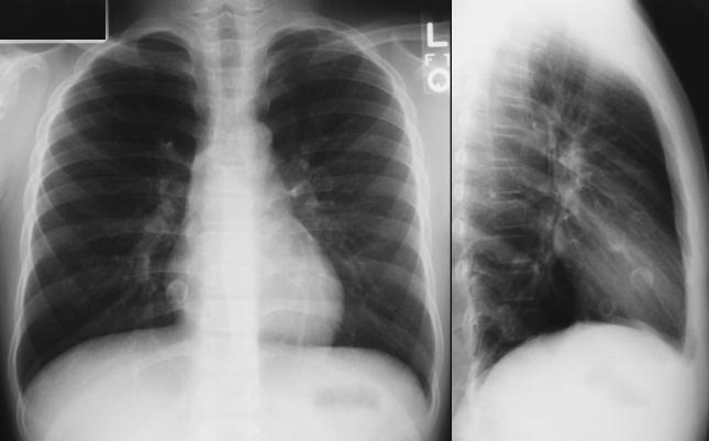 PA and lateral views of the chest are shown here.
You must enlarge the image to appreciate the findings
here. The heart size is normal. There are no
pulmonary infiltrates. There are several spherical
calcifications with central lucencies overlying the heart
measuring up to 1.8 cm in size. There are at least four
of these clearly visible on the lateral view overlying the
heart and possibly two more. The PA view shows one
of these clearly adjacent to the right inferior heart
border.
A translator is arranged on a three-way telephone
translation access line so that more history can be
obtained. His parents indicate that he had a severe
case of Kawasaki disease when he was two years old.
(10 years ago). During his hospitalization, he
developed heart failure. After his hospitalization, he
had to take heart medicines and aspirin at home. He
sees a heart specialist at home who examines him
twice a year. He last had a chest radiograph one-year
ago. His parents give you the name and phone number
of his cardiologist.
With the translator still on the line, a phone call to
his cardiologist across several time zones is successful.
The cardiologist confirms his past history of Kawasaki
disease. The child developed severe coronary
aneurysms and congestive heart failure at age 2 years.
IV gamma globulin therapy that is used today to reduce
the likelihood of developing coronary aneurysms, was
not in use at the time of his initial illness 10 years ago.
He is now followed periodically. He no longer requires
medications for congestive heart failure. You describe
the spherical pearl-like calcifications on his chest
radiograph. The cardiologist indicates that this is
nothing new since these have been visible on his chest
radiographs for many years now. These represent
calcifications of his coronary aneurysms.
Some the clinical manifestations of Kawasaki
disease are described in Case 1 of Volume 3,
Myocardial Failure in a 2-Month Old. Coronary
aneurysms are a known complication of Kawasaki
disease. Acutely, coronary aneurysms may thrombose
resulting in coronary insufficiency. Myocarditis may
also develop resulting in cardiogenic congestive heart
failure and/or shock. Cardiogenic shock in young
children may present with vomiting. While vomiting is
often assumed to be due to viral gastrointestinal
infections, a careful assessment of perfusion
parameters and cardiac auscultation should prompt the
physician to consider cardiac conditions. Myocarditis
may often present with muffled heart tones. Thus, it is
important to ascertain the integrity of the heart tones in
children presenting with vomiting or other symptoms
suggestive of congestive heart failure.
During the years following the acute phase of
Kawasaki disease, small aneurysms will usually resolve
without complications. Others may evolve resulting in
coronary vessel stenosis subjecting such patients to an
increased risk of myocardial ischemia and infarction in
later life. Large coronary calcifications such as the
ones seen on this patient's chest radiograph are
unusual. This case is useful to appreciate the
magnitude of coronary vessel damage in some children
with Kawasaki disease. Thus, children or teenagers
with a past history of Kawasaki disease presenting with
chest pain suggestive of ischemia should be treated as
a rule out myocardial infarction since their degree of
coronary vessel disease may be severe.
References
Yamamoto LG, Martin JG. Kawasaki syndrome in
the ED. Am J Emerg. Med 1994;12:178-182.
Melish ME. Hicks RV. Kawasaki Syndrome: Clinical
features, pathophysiology, etiology, and therapy. J
Rheumatoloty (suppl 24) `1990;17:2-10.
Gersony WM. Diagnosis and management of
Kawasaki disease. JAMA 1991;256(20):2699-2703.
PA and lateral views of the chest are shown here.
You must enlarge the image to appreciate the findings
here. The heart size is normal. There are no
pulmonary infiltrates. There are several spherical
calcifications with central lucencies overlying the heart
measuring up to 1.8 cm in size. There are at least four
of these clearly visible on the lateral view overlying the
heart and possibly two more. The PA view shows one
of these clearly adjacent to the right inferior heart
border.
A translator is arranged on a three-way telephone
translation access line so that more history can be
obtained. His parents indicate that he had a severe
case of Kawasaki disease when he was two years old.
(10 years ago). During his hospitalization, he
developed heart failure. After his hospitalization, he
had to take heart medicines and aspirin at home. He
sees a heart specialist at home who examines him
twice a year. He last had a chest radiograph one-year
ago. His parents give you the name and phone number
of his cardiologist.
With the translator still on the line, a phone call to
his cardiologist across several time zones is successful.
The cardiologist confirms his past history of Kawasaki
disease. The child developed severe coronary
aneurysms and congestive heart failure at age 2 years.
IV gamma globulin therapy that is used today to reduce
the likelihood of developing coronary aneurysms, was
not in use at the time of his initial illness 10 years ago.
He is now followed periodically. He no longer requires
medications for congestive heart failure. You describe
the spherical pearl-like calcifications on his chest
radiograph. The cardiologist indicates that this is
nothing new since these have been visible on his chest
radiographs for many years now. These represent
calcifications of his coronary aneurysms.
Some the clinical manifestations of Kawasaki
disease are described in Case 1 of Volume 3,
Myocardial Failure in a 2-Month Old. Coronary
aneurysms are a known complication of Kawasaki
disease. Acutely, coronary aneurysms may thrombose
resulting in coronary insufficiency. Myocarditis may
also develop resulting in cardiogenic congestive heart
failure and/or shock. Cardiogenic shock in young
children may present with vomiting. While vomiting is
often assumed to be due to viral gastrointestinal
infections, a careful assessment of perfusion
parameters and cardiac auscultation should prompt the
physician to consider cardiac conditions. Myocarditis
may often present with muffled heart tones. Thus, it is
important to ascertain the integrity of the heart tones in
children presenting with vomiting or other symptoms
suggestive of congestive heart failure.
During the years following the acute phase of
Kawasaki disease, small aneurysms will usually resolve
without complications. Others may evolve resulting in
coronary vessel stenosis subjecting such patients to an
increased risk of myocardial ischemia and infarction in
later life. Large coronary calcifications such as the
ones seen on this patient's chest radiograph are
unusual. This case is useful to appreciate the
magnitude of coronary vessel damage in some children
with Kawasaki disease. Thus, children or teenagers
with a past history of Kawasaki disease presenting with
chest pain suggestive of ischemia should be treated as
a rule out myocardial infarction since their degree of
coronary vessel disease may be severe.
References
Yamamoto LG, Martin JG. Kawasaki syndrome in
the ED. Am J Emerg. Med 1994;12:178-182.
Melish ME. Hicks RV. Kawasaki Syndrome: Clinical
features, pathophysiology, etiology, and therapy. J
Rheumatoloty (suppl 24) `1990;17:2-10.
Gersony WM. Diagnosis and management of
Kawasaki disease. JAMA 1991;256(20):2699-2703.
Return to Radiology Cases In Ped Emerg Med Case Selection Page
Return to Univ. Hawaii Dept. Pediatrics Home Page