Sunrise View of the Knee
Radiology Cases in Pediatric Emergency Medicine
Volume 6, Case 8
Loren G. Yamamoto, MD, MPH
Kapiolani Medical Center For Women And Children
University of Hawaii John A. Burns School of Medicine
This is a 10 year old female who fell onto her knee
on the school playground during recess. She jumped
for some horizontal bars which slipped from her grasp.
She fell onto a hard dirt surface injuring her left knee.
She is able to walk, but this is painful.
Her past medical history is unremarkable.
Exam: VS T36.9 (oral), P1000, R18, BP 110/65.
She is comfortable. Her exam findings are only
significant for the left knee. She points to the medial
side of her patella as the site of pain. There is mild
swelling noted around this region. No abrasions,
lacerations or bruises are noted. The patella is tender.
Range of motion is fair with moderately severe pain.
There is no other deformities. The drawer sign is
negative and lateral stability about the knee is good.
Her tibia, fibula, femur and hip are all non-tender.
Radiographs of her knee are obtained.
View knee radiographs.
View AP.
 View lateral.
 Where do you suspect her to have a fracture? Can
you see this area adequately on an AP and lateral view
of the knee?
Since she fell onto her patella and the site of her
pain and tenderness is limited to the patella, it is very
likely that she has a patella fracture. The patella
cannot be viewed very well on an AP view because the
distal femur overlaps it. Since the distal femur is much
thicker than the patella, it is difficult to see any bony
detail of the patella on AP radiographs of the knee.
The lateral view of the knee isolates the patella well,
but this view will only detect horizontal fractures of the
patella. Vertical or oblique fractures will often not be
visible on a lateral view because the width of the
patella will obscure bony detail in the oblique and
vertical planes.
Another view of the knee is obtained.
View sunrise view.
Where do you suspect her to have a fracture? Can
you see this area adequately on an AP and lateral view
of the knee?
Since she fell onto her patella and the site of her
pain and tenderness is limited to the patella, it is very
likely that she has a patella fracture. The patella
cannot be viewed very well on an AP view because the
distal femur overlaps it. Since the distal femur is much
thicker than the patella, it is difficult to see any bony
detail of the patella on AP radiographs of the knee.
The lateral view of the knee isolates the patella well,
but this view will only detect horizontal fractures of the
patella. Vertical or oblique fractures will often not be
visible on a lateral view because the width of the
patella will obscure bony detail in the oblique and
vertical planes.
Another view of the knee is obtained.
View sunrise view.
 This is called the sunrise view because the patella
appears to be rising over the horizon. This view is
taken with the knee flexed. The radiograph is taken
with the x-ray beam tangential to the patella parallel to
the long axis of the lower extremity. Note the small
avulsion fracture of the medial aspect of the patella
This fracture is in the vertical plane making it difficult to
see on the lateral view of the patella. The sunrise view
helps to identify vertical fractures of the patella.
Whenever a fracture of the patella is suspected, a
sunrise view should be requested. There are many
different views of the knee and the sunrise view is not
considered part of the standard set of views in many
hospitals. It must be specifically ordered.
This second set of radiographs demonstrates the
opposite phenomenon. This is an older patient with
patellar pain and tenderness.
View knee radiographs.
View AP.
This is called the sunrise view because the patella
appears to be rising over the horizon. This view is
taken with the knee flexed. The radiograph is taken
with the x-ray beam tangential to the patella parallel to
the long axis of the lower extremity. Note the small
avulsion fracture of the medial aspect of the patella
This fracture is in the vertical plane making it difficult to
see on the lateral view of the patella. The sunrise view
helps to identify vertical fractures of the patella.
Whenever a fracture of the patella is suspected, a
sunrise view should be requested. There are many
different views of the knee and the sunrise view is not
considered part of the standard set of views in many
hospitals. It must be specifically ordered.
This second set of radiographs demonstrates the
opposite phenomenon. This is an older patient with
patellar pain and tenderness.
View knee radiographs.
View AP.
 View lateral.
 View sunrise.
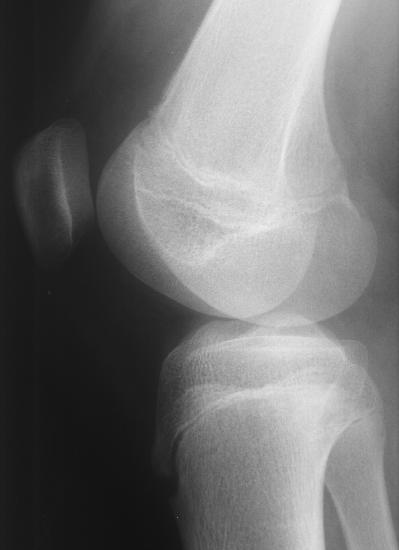
 Again, the AP view does not reveal much because
the patella fracture is obscured by the distal femur.
The lateral view shows the patella fracture well
because the fracture is in the horizontal plane. In this
case, the sunrise view does not show anything because
the fracture is in the horizontal plane (not the vertical
plane).
Despite this fracture being very large, it is not
possible to see it if viewing it in the wrong plane. Even
a large vertical fracture of the patella may not be
visible on the AP and lateral views. A sunrise view will
need to be ordered to visualize the fracture. Similarly,
the patella fracture may occasionally be oblique in
which case all the views may fail to show it well.
Patellar fractures are generally caused by direct
trauma to the patella or due to an avulsion injury of the
quadriceps or patella tendon insertions. Comparison
views may be useful to identify the correct position of
the patella. In a quadriceps or patellar tendon avulsion,
the patella will often be out of position.
A "bipartite" patella is a normal variant that
resembles a patella fracture. A comparison view may
be helpful to distinguish this from a fracture. The
lucency in a bipartite patella is usually in the superior
lateral portion of the patella.
References:
The Patella (Chapter 19). In: Simon RR,
Koenigsknecht SJ. Emergency Orthopedics: The
Extremities, thid edition. 1995, Norwalk, CT, Appleton
& Lange, pp. 287-289.
Again, the AP view does not reveal much because
the patella fracture is obscured by the distal femur.
The lateral view shows the patella fracture well
because the fracture is in the horizontal plane. In this
case, the sunrise view does not show anything because
the fracture is in the horizontal plane (not the vertical
plane).
Despite this fracture being very large, it is not
possible to see it if viewing it in the wrong plane. Even
a large vertical fracture of the patella may not be
visible on the AP and lateral views. A sunrise view will
need to be ordered to visualize the fracture. Similarly,
the patella fracture may occasionally be oblique in
which case all the views may fail to show it well.
Patellar fractures are generally caused by direct
trauma to the patella or due to an avulsion injury of the
quadriceps or patella tendon insertions. Comparison
views may be useful to identify the correct position of
the patella. In a quadriceps or patellar tendon avulsion,
the patella will often be out of position.
A "bipartite" patella is a normal variant that
resembles a patella fracture. A comparison view may
be helpful to distinguish this from a fracture. The
lucency in a bipartite patella is usually in the superior
lateral portion of the patella.
References:
The Patella (Chapter 19). In: Simon RR,
Koenigsknecht SJ. Emergency Orthopedics: The
Extremities, thid edition. 1995, Norwalk, CT, Appleton
& Lange, pp. 287-289.
Return to Radiology Cases In Ped Emerg Med Case Selection Page
Return to Univ. Hawaii Dept. Pediatrics Home Page