Appendicoliths
Radiology Cases in Pediatric Emergency Medicine
Volume 6, Case 18
Loren G. Yamamoto, MD, MPH
Kapiolani Medical Center For Women And Children
University of Hawaii John A. Burns School of Medicine
Collin S. Goto, MD
Children's Medical Center of Dallas
University of Texas Southwestern School of Medicine
Recognizing an appendicolith (fecalith) on abdominal
radiographs can be difficult. Since the presence of an
appendicolith on abdominal radiographs is highly
indicative of appendicitis, identifying a subtle
appendicolith is critical. These 16 cases of radiographic
appendicoliths demonstrate how difficult it is to identify
these reliably. See if you can identify the
appendicoliths in all these radiographs.
For each case, the first image shows the abdominal
films. The second image displays a close up view of
the right lower quadrant. The third image displays
pointers identifying the appendicolith(s).
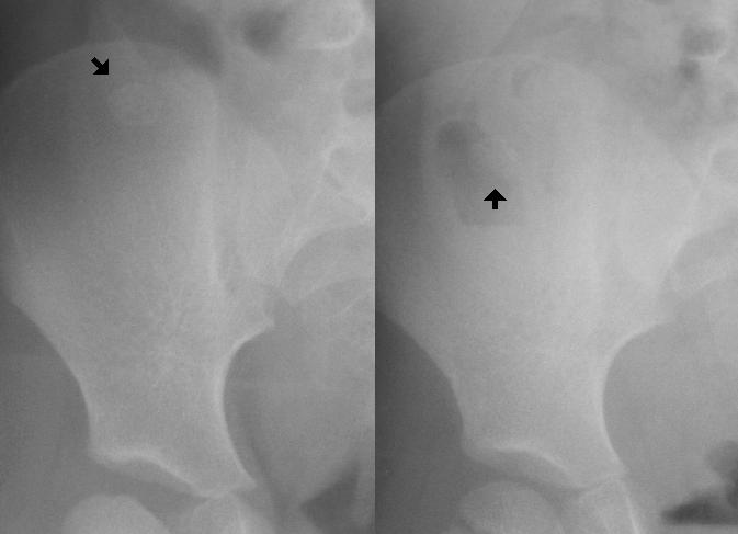
Case A - 7 year old female.
 View close up view of right lower quadrant.
 View pointers.
 Case B - 17 year old female.
 View close up view of right lower quadrant.
 View pointers.
 Case C - 2-1/2 year old male.
 View close up view of right lower quadrant.
 View pointers.
 Case D - 9 year old obese female with a retrocecal
appendicitis.
Case D - 9 year old obese female with a retrocecal
appendicitis.
 View close up view of right lower quadrant.
 View pointers.
 Case E - 8 year old female (supine view only).
 View close up view of right lower quadrant.
 View pointers.
 Case F - 8 year old male (supine and prone views only).
 View close up view of right lower quadrant.
 View pointers.
 Case G - 14 year old female (1 view only). This image
shows a radiograph of the appendix surgical specimen
containing the appendicolith.
Case G - 14 year old female (1 view only). This image
shows a radiograph of the appendix surgical specimen
containing the appendicolith.
 View close up view of right lower quadrant.
 View pointers.
 Case H - 8 year old female.
 View close up view of right lower quadrant.
 View pointers.
 Case I - 9 year old female.
 View close up view of right lower quadrant.
 View pointers.
 Case J - 8 year old male.
 View close up view of right lower quadrant.
 View pointers.
 Case K - Not sure of this patient's age.
 View close up view of right lower quadrant.
 View pointers.
 Case L - Not sure of this patient's age.
 View close up view of right lower quadrant.
 View pointers.
 Case M - 3 year old male.
 View close up view of right lower quadrant.
 View pointers.
 Case N - 6 year old male.
 View close up view of right lower quadrant.
 View pointers.
 Case O - 17 month old female.
 View close up view of right lower quadrant.
 View pointers.
 Case P - 15 year old male.
 View close up view of right lower quadrant.
 View pointers.

Return to Radiology Cases In Ped Emerg Med Case Selection Page
Return to Univ. Hawaii Dept. Pediatrics Home Page