Galeazzi's Injury
Radiology Cases in Pediatric Emergency Medicine
Volume 1, Case 16
Loren G. Yamamoto, MD, MPH
Stanley M.K. Chung, MD
Kapiolani Medical Center For Women And Children
University of Hawaii John A. Burns School of Medicine
A 12 year old male is brought to the ED after
injuring his forearm rollerblading. He fell onto his
palm and noted pain and a deformity in his forearm.
Examination revealed normal vital signs and findings
limited to his left arm. His clavicle, shoulder, humerus,
and hand were non-tender. He was reluctant to move
his shoulder since his forearm was in a splint and sling.
There was an obvious angulation at the mid-forearm.
He could move all his fingers. No circulatory or sensory
deficits were detected. Radiographs of his forearm
were obtained.
View radiographs.
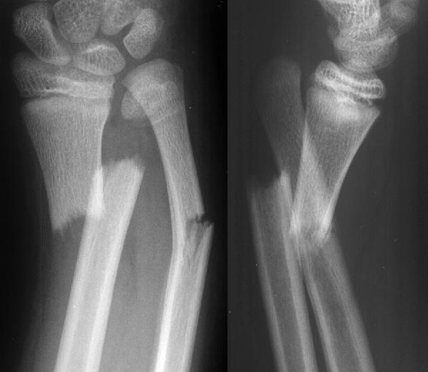 The radiographs show an angulated distal radius
and ulna fracture, a fracture through the physis of the
distal ulna, and a dislocation of the distal ulna
(radioulnar dislocation). What type of Salter-Harris
fracture is present at the distal ulna? If you have
difficulty with the Salter-Harris classification, review
Case 18 (Salter-Harris). This is probably a SH type I
fracture, although small parts of the metaphysis may
still be attached to the fracture segment, making it a
type II.
The classic Galeazzi fracture is described as a
fracture of the distal third of the radius associated with a
dislocation of the distal ulna. This classic injury occurs
more commonly in adults and teenagers than in
younger children. It may be very difficult to recognize
since the radioulnar joint is painful and difficult to
examine in the presence of an adjacent radius fracture.
The radioulnar joint may spontaneously reduce in some
instances. Orthopedic surgeons usually examine the
radioulnar joint stability during reduction of the radius
fracture or by using more advanced imaging methods.
One should be suspicious of the Galeazzi fracture in
any angulated fracture of the distal radius. Radioulnar
dislocation is unlikely in simple non-angulated
torus-type distal radius fractures.
A Galeazzi-like injury occurs in children. The
ligaments of the distal forearm normally prevent the
radius and ulna from twisting about each other. As the
distal radius fractures, exaggerated twisting forces in
the hyperpronation or hypersupination direction result in
the loss of stabilization of the radioulnar attachments.
Ligaments attaching the distal ulnar epiphysis to the
distal radius and the carpal/metacarpals gain tension on
the ulnar head as twisting occurs. If the tension force is
great enough, the ulnar physis fractures. Recognizing a
Salter-Harris type I or II fracture of the ulnar physis
associated with a distal radius fracture should cause
one to consider the possible complications of th
e Galeazzi fracture.
If the Galeazzi injury is not recognized, the
radioulnar dislocation may not be identified. This can
result in a painful prominence of the distal ulna.
Occasionally, the extensor digitorum communis tendon
may become entrapped between the ulnar epiphysis
and metaphysis in children, making closed reduction
impossible.
References
Letts RM. Monteggia and Galeazzi Fractures. In:
Letts RM (ed): Management of Pediatric Fractures.
New York, Churchill Livingstone, 1994, pp. 295-321.
The radiographs show an angulated distal radius
and ulna fracture, a fracture through the physis of the
distal ulna, and a dislocation of the distal ulna
(radioulnar dislocation). What type of Salter-Harris
fracture is present at the distal ulna? If you have
difficulty with the Salter-Harris classification, review
Case 18 (Salter-Harris). This is probably a SH type I
fracture, although small parts of the metaphysis may
still be attached to the fracture segment, making it a
type II.
The classic Galeazzi fracture is described as a
fracture of the distal third of the radius associated with a
dislocation of the distal ulna. This classic injury occurs
more commonly in adults and teenagers than in
younger children. It may be very difficult to recognize
since the radioulnar joint is painful and difficult to
examine in the presence of an adjacent radius fracture.
The radioulnar joint may spontaneously reduce in some
instances. Orthopedic surgeons usually examine the
radioulnar joint stability during reduction of the radius
fracture or by using more advanced imaging methods.
One should be suspicious of the Galeazzi fracture in
any angulated fracture of the distal radius. Radioulnar
dislocation is unlikely in simple non-angulated
torus-type distal radius fractures.
A Galeazzi-like injury occurs in children. The
ligaments of the distal forearm normally prevent the
radius and ulna from twisting about each other. As the
distal radius fractures, exaggerated twisting forces in
the hyperpronation or hypersupination direction result in
the loss of stabilization of the radioulnar attachments.
Ligaments attaching the distal ulnar epiphysis to the
distal radius and the carpal/metacarpals gain tension on
the ulnar head as twisting occurs. If the tension force is
great enough, the ulnar physis fractures. Recognizing a
Salter-Harris type I or II fracture of the ulnar physis
associated with a distal radius fracture should cause
one to consider the possible complications of th
e Galeazzi fracture.
If the Galeazzi injury is not recognized, the
radioulnar dislocation may not be identified. This can
result in a painful prominence of the distal ulna.
Occasionally, the extensor digitorum communis tendon
may become entrapped between the ulnar epiphysis
and metaphysis in children, making closed reduction
impossible.
References
Letts RM. Monteggia and Galeazzi Fractures. In:
Letts RM (ed): Management of Pediatric Fractures.
New York, Churchill Livingstone, 1994, pp. 295-321.
Return to Radiology Cases In Ped Emerg Med Case Selection Page
Return to Univ. Hawaii Dept. Pediatrics Home Page