Periumbilical Abdominal Pain
Radiology Cases in Pediatric Emergency Medicine
Volume 4, Case 9
Loren G. Yamamoto, MD, MPH
Kapiolani Medical Center For Women And Children
University of Hawaii John A. Burns School of Medicine
This is a 10-year old male presenting to the E.D.
with a history of abdominal pain for two days. He
describes the pain mostly in his periumbilical region.
The pain is clearly worse today. He has no vomiting or
diarrhea. His appetite is poor and he is not able to
ambulate well due to pain. There is no history of fever.
There is no history of coughing, chest pain, or dysuria.
His past history is significant for asthma.
Exam: VS T37.4 (tympanic), P92, R16, BP 120/62.
He is alert, not toxic, resting comfortably. His hydration
is good. Heart regular without murmurs. Lungs clear.
Abdomen is flat and generally soft. There is mild
guarding and tenderness mostly over the periumbilical
region. Bowel sounds are absent. No hernias are
evident. Observing his gait, he ambulates slowly in a
bent forward position. He refuses to jump. Asking him
to cough results in moderately severe abdominal pain.
A rectal exam does not yield any localizing signs.
Laboratory studies: CBC WBC 14,500, 81% segs,
2% bands, 8% lymphs, 6% monos, 3% atypical lymphs,
Hgb. 14.3, Hct. 43.0, platelet count 344,000. Urinalysis
SG 1.030, trace protein, otherwise negative.
An abdominal series is obtained.
View abdominal series: Flat (supine) view.
 View upright view.
 Given the patient's clinical findings, consider the
differential diagnosis at this point and what we should
be most interested in, in examining these radiographs.
For example, an obstruction is not likely given the
absence of previous abdominal surgery, the absence of
vomiting, the flat abdominal contour, and no clinical
evidence of an incarcerated hernia. Appendicitis is a
consideration given the peritoneal signs exhibited, the
patient's gait suggesting peritonitis, his anorexia, and
modest leukocytosis. His pain and tenderness are not
in the right lower quadrant. However, the absence of
this cannot reliably exclude appendicitis.
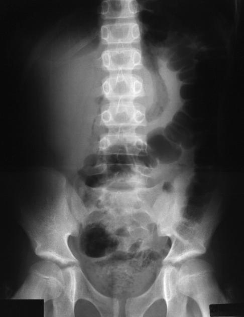
These films are dark; making the lateral edges of the
abdomen difficult to view. The gas distribution shows
gas and feces throughout the colon. However, the
ascending colon is displaced medially. It appears to be
separated from the right flank, raising the possibility of a
mass lateral to the colon. There is no bowel dilation
and no air fluid levels. No fecolith is seen. No free air
is evident.
An abdominal ultrasound is performed. There is
evidence of a fluid and gas-containing structure
adjacent to the umbilicus consistent with an ectopic
appendicitis or a Meckel's diverticulitis.
An exploratory laparotomy is performed. An acutely
inflamed Meckel's diverticulum is noted. This is
resected. His appendix is normal. He is placed on
antibiotics post-operatively, and he recovers
uneventfully.
The radiographic findings of appendicitis are highly
variable. This is discussed in some detail in Case 19 of
Volume 3, Abdominal Pain and the Peritoneal Fat
Margins. This case specifically discusses the
significance of the peritoneal fat margins. Usually, the
bowel is close to the peritoneal fat margins bilaterally,
but in this current case, the bowel is displaced far from
the right peritoneal fat margin displaying a mass effect.
These radiographs were too dark for the scanner to
pick-up the peritoneal fat margins on the image
displayed here. However, since this case is extreme, it
is evident that the ascending colon is being displaced
medially. Often this displacement of the bowel away
from the peritoneal fat margin can be subtle. Although
this patient's diagnosis is an unusual one, the general
principles of identifying a patient requiring prompt
abdominal surgery still apply.
Given the patient's clinical findings, consider the
differential diagnosis at this point and what we should
be most interested in, in examining these radiographs.
For example, an obstruction is not likely given the
absence of previous abdominal surgery, the absence of
vomiting, the flat abdominal contour, and no clinical
evidence of an incarcerated hernia. Appendicitis is a
consideration given the peritoneal signs exhibited, the
patient's gait suggesting peritonitis, his anorexia, and
modest leukocytosis. His pain and tenderness are not
in the right lower quadrant. However, the absence of
this cannot reliably exclude appendicitis.
These films are dark; making the lateral edges of the
abdomen difficult to view. The gas distribution shows
gas and feces throughout the colon. However, the
ascending colon is displaced medially. It appears to be
separated from the right flank, raising the possibility of a
mass lateral to the colon. There is no bowel dilation
and no air fluid levels. No fecolith is seen. No free air
is evident.
An abdominal ultrasound is performed. There is
evidence of a fluid and gas-containing structure
adjacent to the umbilicus consistent with an ectopic
appendicitis or a Meckel's diverticulitis.
An exploratory laparotomy is performed. An acutely
inflamed Meckel's diverticulum is noted. This is
resected. His appendix is normal. He is placed on
antibiotics post-operatively, and he recovers
uneventfully.
The radiographic findings of appendicitis are highly
variable. This is discussed in some detail in Case 19 of
Volume 3, Abdominal Pain and the Peritoneal Fat
Margins. This case specifically discusses the
significance of the peritoneal fat margins. Usually, the
bowel is close to the peritoneal fat margins bilaterally,
but in this current case, the bowel is displaced far from
the right peritoneal fat margin displaying a mass effect.
These radiographs were too dark for the scanner to
pick-up the peritoneal fat margins on the image
displayed here. However, since this case is extreme, it
is evident that the ascending colon is being displaced
medially. Often this displacement of the bowel away
from the peritoneal fat margin can be subtle. Although
this patient's diagnosis is an unusual one, the general
principles of identifying a patient requiring prompt
abdominal surgery still apply.
Return to Radiology Cases In Ped Emerg Med Case Selection Page
Return to Univ. Hawaii Dept. Pediatrics Home Page