More Cervical Spine Injuries
Radiology Cases in Pediatric Emergency Medicine
Volume 5, Case 5
Loren G. Yamamoto, MD, MPH
Kapiolani Medical Center For Women And Children
University of Hawaii John A. Burns School of Medicine
Test your skill in reading these 16 pediatric cervical
spine radiographs. Many of these have subtle findings.
Follow the principles outlined in Case 2 of this volume,
Cervical Spine Radiographs, and see how accurate
you can be at interpreting these radiographs.
View Case A
 Interpretation of Case A
Lateral and AP views are shown here. The top of
the lateral view was cut off, thus visualizing only part of
C1. There is an obvious compression fracture of C5.
C4 and C7 are also compressed. There is an avulsion
of the anterior superior lip of C7.
The AP view shows a vertical fracture through C5.
Impression: Compression fractures of C4, C5, C7.
View Case B
Interpretation of Case A
Lateral and AP views are shown here. The top of
the lateral view was cut off, thus visualizing only part of
C1. There is an obvious compression fracture of C5.
C4 and C7 are also compressed. There is an avulsion
of the anterior superior lip of C7.
The AP view shows a vertical fracture through C5.
Impression: Compression fractures of C4, C5, C7.
View Case B
 Interpretation of Case B
Two lateral views are shown here. Both radiographs
show that C4 is displaced anteriorly with respect to C5.
The anterior vertebral body line, the posterior vertebral
body (anterior spinal canal) line, and the spinolaminal
(posterior spinal canal) line are out of alignment.
Impression: C4-C5 subluxation.
View Case C
Radiographs contributed by Martin I. Herman, MD
Interpretation of Case B
Two lateral views are shown here. Both radiographs
show that C4 is displaced anteriorly with respect to C5.
The anterior vertebral body line, the posterior vertebral
body (anterior spinal canal) line, and the spinolaminal
(posterior spinal canal) line are out of alignment.
Impression: C4-C5 subluxation.
View Case C
Radiographs contributed by Martin I. Herman, MD
 Interpretation of Case C
This lateral view shows C4 slightly displaced
anteriorly with respect to C5. Someone has placed a
strip of tape over the arch of C5 to show that the
posterior border of the C5 vertebral body does not line
up with the other vertebral bodies. The borders of the
tape introduce some artifact. The lucency that appears
over the arch of C6 is from the tape (not a fracture).
The anterior vertebral body line, the posterior vertebral
body (anterior spinal canal) line, and the spinolaminal
(posterior spinal canal) line are out of alignment.
Impression: C4-C5 subluxation.
View Case D
Radiographs contributed by Martin I. Herman, MD
Interpretation of Case C
This lateral view shows C4 slightly displaced
anteriorly with respect to C5. Someone has placed a
strip of tape over the arch of C5 to show that the
posterior border of the C5 vertebral body does not line
up with the other vertebral bodies. The borders of the
tape introduce some artifact. The lucency that appears
over the arch of C6 is from the tape (not a fracture).
The anterior vertebral body line, the posterior vertebral
body (anterior spinal canal) line, and the spinolaminal
(posterior spinal canal) line are out of alignment.
Impression: C4-C5 subluxation.
View Case D
Radiographs contributed by Martin I. Herman, MD
 Interpretation of Case D
This lateral view shows C2 tilted anteriorly over C3.
While one might consider the possibility that this is a
C2-C3 pseudosubluxation, this degree of angulation is
excessive. Additionally, the C2-C3 facet joints are
disrupted.
Radiographic features consistent with a C2-C3
pseudosubluxation are:
1) Neck position should be neutral or in flexion.
However, in the case of this radiograph, the lower
portion of the neck (C3 to C7) is in extension (lordosis).
The only flexion in the neck is at the C2-C3 region,
which is abnormal. Since this criterion is not met, this is
not consistent with a pseudosubluxation.
2) The Swischuk line should be in good alignment.
This is a line drawn from the anterior aspect of the
posterior arch of C1 to the anterior aspect of the
posterior arch of C3. The anterior aspect of the
posterior arch of C2 should be within 1.5 mm of this line
(refer to Case 3 of this volume and Case 5 of Volume
1). In the case of this radiograph, the Swischuk line
alignment is satisfactory.
3) Other factors favoring a pseudosubluxation
include a benign mechanism of injury, low clinical risk,
and resolution of the pseudosubluxation upon repeating
the radiograph following repositioning the neck in
extension (lordosis) (This often cannot be done if a true
subluxation is suspected).
Thus, a satisfactory alignment of the Swischuk line
alone is not sufficient to rule out a true subluxation.
The anterior vertebral body line, the posterior
vertebral body (anterior spinal canal) line, and the
spinolaminal (posterior spinal canal) line are out of
alignment.
Impression: C2-C3 subluxation.
View Case E
Interpretation of Case D
This lateral view shows C2 tilted anteriorly over C3.
While one might consider the possibility that this is a
C2-C3 pseudosubluxation, this degree of angulation is
excessive. Additionally, the C2-C3 facet joints are
disrupted.
Radiographic features consistent with a C2-C3
pseudosubluxation are:
1) Neck position should be neutral or in flexion.
However, in the case of this radiograph, the lower
portion of the neck (C3 to C7) is in extension (lordosis).
The only flexion in the neck is at the C2-C3 region,
which is abnormal. Since this criterion is not met, this is
not consistent with a pseudosubluxation.
2) The Swischuk line should be in good alignment.
This is a line drawn from the anterior aspect of the
posterior arch of C1 to the anterior aspect of the
posterior arch of C3. The anterior aspect of the
posterior arch of C2 should be within 1.5 mm of this line
(refer to Case 3 of this volume and Case 5 of Volume
1). In the case of this radiograph, the Swischuk line
alignment is satisfactory.
3) Other factors favoring a pseudosubluxation
include a benign mechanism of injury, low clinical risk,
and resolution of the pseudosubluxation upon repeating
the radiograph following repositioning the neck in
extension (lordosis) (This often cannot be done if a true
subluxation is suspected).
Thus, a satisfactory alignment of the Swischuk line
alone is not sufficient to rule out a true subluxation.
The anterior vertebral body line, the posterior
vertebral body (anterior spinal canal) line, and the
spinolaminal (posterior spinal canal) line are out of
alignment.
Impression: C2-C3 subluxation.
View Case E
 Interpretation of Case E
This lateral view shows C2 slightly displaced
anteriorly with respect to C3. The anterior vertebral
body line, the posterior vertebral body (anterior spinal
canal) line, and the spinolaminal (posterior spinal canal)
line, are out of alignment. This radiograph shows poor
positioning. It is not a true lateral, rather it is oblique.
Note the prominence of the intervertebral foramina
which are most prominent on an oblique view.
Unlike case D, this radiograph shows the entire
cervical spine to be in flexion. The Swischuk line is at
the limits of tolerance in this case since the anterior
aspect of the posterior arch of C2 is about 1.5 mm from
the Swischuk line; however it is not a true lateral view.
Clinically, this patient's mechanism of injury is low
risk and her degree of discomfort is felt to be most
consistent with a pseudosubluxation. Repeat films of
her neck in better positioning are normal.
Impression: Probable C2-C3 pseudosubluxation.
The anterior vertebral body line, the posterior vertebral
body (anterior spinal canal) line, and the spinolaminal
(posterior spinal canal) line, are out of alignment
probably due to poor positioning.
View Case F
Radiographs contributed by Collin S. Goto, MD
Interpretation of Case E
This lateral view shows C2 slightly displaced
anteriorly with respect to C3. The anterior vertebral
body line, the posterior vertebral body (anterior spinal
canal) line, and the spinolaminal (posterior spinal canal)
line, are out of alignment. This radiograph shows poor
positioning. It is not a true lateral, rather it is oblique.
Note the prominence of the intervertebral foramina
which are most prominent on an oblique view.
Unlike case D, this radiograph shows the entire
cervical spine to be in flexion. The Swischuk line is at
the limits of tolerance in this case since the anterior
aspect of the posterior arch of C2 is about 1.5 mm from
the Swischuk line; however it is not a true lateral view.
Clinically, this patient's mechanism of injury is low
risk and her degree of discomfort is felt to be most
consistent with a pseudosubluxation. Repeat films of
her neck in better positioning are normal.
Impression: Probable C2-C3 pseudosubluxation.
The anterior vertebral body line, the posterior vertebral
body (anterior spinal canal) line, and the spinolaminal
(posterior spinal canal) line, are out of alignment
probably due to poor positioning.
View Case F
Radiographs contributed by Collin S. Goto, MD
 Interpretation of Case F
This is an 18-month old male riding unrestrained in
the front passenger seat of a car involved in a motor
vehicle collision. He was ejected from the vehicle,
sustaining multiple trauma.
Lateral and AP views are shown here. The lateral
view shows separation of the skull from the cervical
spine (atlanto-occipital dislocation).
NOTE: There is a visible lucency at the base of the
odontoid. This is the subdental synchondrosis, a
normal finding in young children. This synchondrosis
generally fuses by age 3 to 6 years.
Impression: Atlanto-occipital dislocation.
View Case G
Radiographs contributed by Collin S. Goto, MD
Interpretation of Case F
This is an 18-month old male riding unrestrained in
the front passenger seat of a car involved in a motor
vehicle collision. He was ejected from the vehicle,
sustaining multiple trauma.
Lateral and AP views are shown here. The lateral
view shows separation of the skull from the cervical
spine (atlanto-occipital dislocation).
NOTE: There is a visible lucency at the base of the
odontoid. This is the subdental synchondrosis, a
normal finding in young children. This synchondrosis
generally fuses by age 3 to 6 years.
Impression: Atlanto-occipital dislocation.
View Case G
Radiographs contributed by Collin S. Goto, MD
 Interpretation of Case G
This is a 5-year old female who fell off a trampoline
onto her head with her neck flexed. She presented to
the E.D. with neck pain and tingling in her feet.
Two lateral views are shown here. The anterior
vertebral body line, the posterior vertebral body
(anterior spinal canal) line, and the spinolaminal
(posterior spinal canal) line, are out of alignment. C3 is
displaced anteriorly with respect to C4. The facet joints
of C3/C4 are out of alignment. There is possible facet
joint subluxation at C2/C3.
Impression: C3-C4 subluxation.
View Case H
Interpretation of Case G
This is a 5-year old female who fell off a trampoline
onto her head with her neck flexed. She presented to
the E.D. with neck pain and tingling in her feet.
Two lateral views are shown here. The anterior
vertebral body line, the posterior vertebral body
(anterior spinal canal) line, and the spinolaminal
(posterior spinal canal) line, are out of alignment. C3 is
displaced anteriorly with respect to C4. The facet joints
of C3/C4 are out of alignment. There is possible facet
joint subluxation at C2/C3.
Impression: C3-C4 subluxation.
View Case H
 Interpretation of Case H
A lateral view is shown here. The pre-vertebral soft
tissue space is widened suggesting the possibility of
hemorrhage into this area from a fracture. An NG tube
is in place. An NG tube in the esophagus could widen
the pre-vertebral soft tissue space as well. In this
instance, it is not certain if the widening of the
pre-vertebral soft tissue space is pathologic.
The anterior vertebral body line, the spinolaminal
(posterior spinal canal) line, and the spinous processes
tips line are all in satisfactory alignment. The posterior
vertebral body line is slightly disrupted at the C6-C7
junction where C6 appears to be displaced slightly
anterior with respect to C7 (difficult to see). The
anterior vertebral body line may also be slightly
disrupted at C6-C7, however, this is so slight that it is
difficult to be certain. There is a possible irregularity of
the posterior inferior corner of the C6 vertebral body.
This is possibly a small avulsion fracture. The facets of
C6 are displaced slightly anteriorly with respect to the
facets of C7.
Impression: Possible small avulsion fracture of the
posterior inferior corner of the C6 vertebral body.
Possible anterior displacement of the C6 with respect to
C7. These abnormalities are not definite. The study is
possibly normal.
View Case I
Interpretation of Case H
A lateral view is shown here. The pre-vertebral soft
tissue space is widened suggesting the possibility of
hemorrhage into this area from a fracture. An NG tube
is in place. An NG tube in the esophagus could widen
the pre-vertebral soft tissue space as well. In this
instance, it is not certain if the widening of the
pre-vertebral soft tissue space is pathologic.
The anterior vertebral body line, the spinolaminal
(posterior spinal canal) line, and the spinous processes
tips line are all in satisfactory alignment. The posterior
vertebral body line is slightly disrupted at the C6-C7
junction where C6 appears to be displaced slightly
anterior with respect to C7 (difficult to see). The
anterior vertebral body line may also be slightly
disrupted at C6-C7, however, this is so slight that it is
difficult to be certain. There is a possible irregularity of
the posterior inferior corner of the C6 vertebral body.
This is possibly a small avulsion fracture. The facets of
C6 are displaced slightly anteriorly with respect to the
facets of C7.
Impression: Possible small avulsion fracture of the
posterior inferior corner of the C6 vertebral body.
Possible anterior displacement of the C6 with respect to
C7. These abnormalities are not definite. The study is
possibly normal.
View Case I
 Interpretation of Case I
Two lateral views and a single AP view are shown
here. The lateral view on the right shows an obvious
fracture of the odontoid. However, note that on the
other lateral view of the same patient, the odontoid
fracture is not as easy to appreciate. In the lateral view
on the left, the odontoid fracture can be identified by the
angulation of the odontoid. The other bony elements
are normal. Alignment is satisfactory otherwise.
NOTE: The fracture at the base of the odontoid
could possibly be confused with the normal lucency at
the base of the odontoid in young children (the
subdental synchondrosis). However, while it may be
normal for the odontoid to tilt backward (posteriorly), it
should NOT be tilting forward (anteriorly). Anterior
tilting of the odontoid with a widening of the lucency at
the base of the odontoid are highly indicative of a
fracture and not a normal synchondrosis.
Impression: Odontoid fracture.
View Case J
Interpretation of Case I
Two lateral views and a single AP view are shown
here. The lateral view on the right shows an obvious
fracture of the odontoid. However, note that on the
other lateral view of the same patient, the odontoid
fracture is not as easy to appreciate. In the lateral view
on the left, the odontoid fracture can be identified by the
angulation of the odontoid. The other bony elements
are normal. Alignment is satisfactory otherwise.
NOTE: The fracture at the base of the odontoid
could possibly be confused with the normal lucency at
the base of the odontoid in young children (the
subdental synchondrosis). However, while it may be
normal for the odontoid to tilt backward (posteriorly), it
should NOT be tilting forward (anteriorly). Anterior
tilting of the odontoid with a widening of the lucency at
the base of the odontoid are highly indicative of a
fracture and not a normal synchondrosis.
Impression: Odontoid fracture.
View Case J
 Interpretation of Case J
Lateral, AP, and odontoid views are shown here.
C7 is not visualized well on the lateral view, making this
study inadequate. On the lateral view, C2 may be
slightly displaced anteriorly with respect to C3. The
anterior vertebral body line identifies this displacement
best. The posterior vertebral body (anterior spinal
canal) line, and the spinolaminal (posterior spinal canal)
line, are within satisfactory alignment. The lateral view
also shows an irregularity of the anterior inferior corner
of the C2 vertebral body. This can only be seen on the
enlarged view. It resembles a small drop dripping from
the vertebral body (difficult to see). This is a small
avulsion fracture. The pre-vertebral soft tissue space is
within normal limits.
The AP view shows good alignment of the spinous
processes and equal spacing. The odontoid view
shows the lateral masses of C1 well-positioned with
respect to C2.
Impression: Small avulsion fracture of the anterior
inferior corner of the C2 vertebral body. Slight anterior
displacement of C2 with respect to C3.
View Case K
Interpretation of Case J
Lateral, AP, and odontoid views are shown here.
C7 is not visualized well on the lateral view, making this
study inadequate. On the lateral view, C2 may be
slightly displaced anteriorly with respect to C3. The
anterior vertebral body line identifies this displacement
best. The posterior vertebral body (anterior spinal
canal) line, and the spinolaminal (posterior spinal canal)
line, are within satisfactory alignment. The lateral view
also shows an irregularity of the anterior inferior corner
of the C2 vertebral body. This can only be seen on the
enlarged view. It resembles a small drop dripping from
the vertebral body (difficult to see). This is a small
avulsion fracture. The pre-vertebral soft tissue space is
within normal limits.
The AP view shows good alignment of the spinous
processes and equal spacing. The odontoid view
shows the lateral masses of C1 well-positioned with
respect to C2.
Impression: Small avulsion fracture of the anterior
inferior corner of the C2 vertebral body. Slight anterior
displacement of C2 with respect to C3.
View Case K
 Interpretation of Case K
There is a fracture of the spinous process of C7.
The anterior and posterior vertebral body lines are
satisfactory. The spinolaminal line is satisfactory. The
tips of the spinous processes are difficult to see with the
exception of C7, which is fractured.
This radiograph demonstrates a modest degree of
"fanning". Normally, the spinous processes are evenly
spaced and they converge toward a point because of
their attachment by the posterior longitudinal ligament
and the interspinous ligament. However, this
radiograph shows that the spinous process of C7 is not
converging toward the same point as the other spinous
processes. This spreading of the spinous processes,
known as fanning, is consistent with a fracture of the
spinous process or a tear of the posterior longitudinal
ligament.
This has the appearance of a typical "clay
shoveller's" fracture, which generally occurs when the
neck is forced forward (flexed) while it is held in
extension (lordosis). In this case, this teenager was
swimming when someone diving from the rocks above,
fell onto his back.
Impression: C7 spinous process fracture.
View Case L
Interpretation of Case K
There is a fracture of the spinous process of C7.
The anterior and posterior vertebral body lines are
satisfactory. The spinolaminal line is satisfactory. The
tips of the spinous processes are difficult to see with the
exception of C7, which is fractured.
This radiograph demonstrates a modest degree of
"fanning". Normally, the spinous processes are evenly
spaced and they converge toward a point because of
their attachment by the posterior longitudinal ligament
and the interspinous ligament. However, this
radiograph shows that the spinous process of C7 is not
converging toward the same point as the other spinous
processes. This spreading of the spinous processes,
known as fanning, is consistent with a fracture of the
spinous process or a tear of the posterior longitudinal
ligament.
This has the appearance of a typical "clay
shoveller's" fracture, which generally occurs when the
neck is forced forward (flexed) while it is held in
extension (lordosis). In this case, this teenager was
swimming when someone diving from the rocks above,
fell onto his back.
Impression: C7 spinous process fracture.
View Case L
 Interpretation of Case L
Several views are shown here. The upper left image
is a lateral view which only shows C1 to the upper
portion of C5. Two oblique views are shown in the
upper right. The lower left image is an AP view. The
right lower image shows two swimmer's views.
The lateral view show no pre-vertebral soft tissue
widening. C1 to the top of C5 are in satisfactory
alignment. The swimmer's views show poor images of
C5 and C6. C7 is still not well visualized. The anterior
portion of the C6 vertebral body is slightly shorter than
the posterior portion, indicating the possible presence of
a compression fracture. While the height of C6 seen on
the AP and oblique views may seem slightly short, it is
probably within normal limits.
Impression: Possible compression fracture of the
anterior portion of the C6 vertebral body. C7 is not
visualized well.
View Case M
Interpretation of Case L
Several views are shown here. The upper left image
is a lateral view which only shows C1 to the upper
portion of C5. Two oblique views are shown in the
upper right. The lower left image is an AP view. The
right lower image shows two swimmer's views.
The lateral view show no pre-vertebral soft tissue
widening. C1 to the top of C5 are in satisfactory
alignment. The swimmer's views show poor images of
C5 and C6. C7 is still not well visualized. The anterior
portion of the C6 vertebral body is slightly shorter than
the posterior portion, indicating the possible presence of
a compression fracture. While the height of C6 seen on
the AP and oblique views may seem slightly short, it is
probably within normal limits.
Impression: Possible compression fracture of the
anterior portion of the C6 vertebral body. C7 is not
visualized well.
View Case M
 Interpretation of Case M
Multiple views are shown here. The upper left
image is the lateral view. The upper right image is an
odontoid view with the AP view beneath it. The lower
left images are oblique views. The lower right image is
a swimmer's view.
The lateral view shows C1 to the upper portion of
C6. C7 is not visualized. C4 and C5 show
compression fractures of the anterior portions of the C4
and C5 vertebral bodies. C3 appears to be slightly
displaced anteriorly with respect to C4 on both the
lateral view and the swimmer's view.
C2 appears to be slightly displaced anteriorly with
respect to C3. This is due to kyphosis secondary to the
fractures.
The odontoid view shows the odontoid well centered
within C1. However the lateral margins of the lateral
masses cannot be determined from this odontoid view.
Thus, this particular odontoid view is not of satisfactory
quality.
There are no identifiable abnormalities on the
oblique views. The swimmer's view shows C6 better,
but C7 is still not visualized, making this study
suboptimal.
Impression: Compression fractures of C4 and C5.
Unable to visualize C7.
View Case N
Interpretation of Case M
Multiple views are shown here. The upper left
image is the lateral view. The upper right image is an
odontoid view with the AP view beneath it. The lower
left images are oblique views. The lower right image is
a swimmer's view.
The lateral view shows C1 to the upper portion of
C6. C7 is not visualized. C4 and C5 show
compression fractures of the anterior portions of the C4
and C5 vertebral bodies. C3 appears to be slightly
displaced anteriorly with respect to C4 on both the
lateral view and the swimmer's view.
C2 appears to be slightly displaced anteriorly with
respect to C3. This is due to kyphosis secondary to the
fractures.
The odontoid view shows the odontoid well centered
within C1. However the lateral margins of the lateral
masses cannot be determined from this odontoid view.
Thus, this particular odontoid view is not of satisfactory
quality.
There are no identifiable abnormalities on the
oblique views. The swimmer's view shows C6 better,
but C7 is still not visualized, making this study
suboptimal.
Impression: Compression fractures of C4 and C5.
Unable to visualize C7.
View Case N
 Interpretation of Case N
A lateral view is shown here with the neck in a flexed
position. C2 is out of alignment anteriorly with respect
to C3. There is a lucency through the base of the
odontoid. There is widening of the pre-vertebral soft
tissue. The Swischuk line is in satisfactory alignment.
All of these findings are consistent with a
pseudosubluxation due to the positioning of the neck in
flexion. This film was repeated with a better lordotic
extension of the neck. Both the C2-C3
pseudosubluxation and the pre-vertebral soft tissue
widening resolved.
Impression: Pseudosubluxation. Normal subdental
synchondrosis.
View Case O
Interpretation of Case N
A lateral view is shown here with the neck in a flexed
position. C2 is out of alignment anteriorly with respect
to C3. There is a lucency through the base of the
odontoid. There is widening of the pre-vertebral soft
tissue. The Swischuk line is in satisfactory alignment.
All of these findings are consistent with a
pseudosubluxation due to the positioning of the neck in
flexion. This film was repeated with a better lordotic
extension of the neck. Both the C2-C3
pseudosubluxation and the pre-vertebral soft tissue
widening resolved.
Impression: Pseudosubluxation. Normal subdental
synchondrosis.
View Case O
 Interpretation of Case O
There is fusion of several vertebral units. Alignment
appears to be satisfactory, but normal anatomic
landmarks are not present.
Impression: Congenital fusion of adjacent vertebral
bodies.
View Case P
Interpretation of Case O
There is fusion of several vertebral units. Alignment
appears to be satisfactory, but normal anatomic
landmarks are not present.
Impression: Congenital fusion of adjacent vertebral
bodies.
View Case P
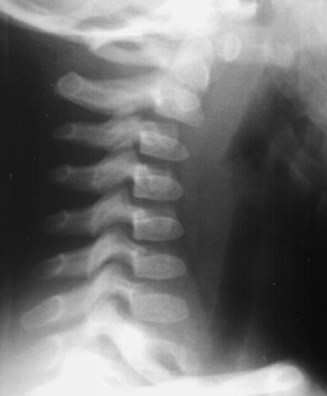
 Interpretation of Case P
C4 and C5 are out of alignment, displaced
posteriorly with respect to the rest of the C-spine. The
anterior aspects of the C4 and C5 vertebral bodies are
shorter than the posterior aspects due to wedge type
compression fractures. The vertebral body of C6 is
shortened due to a compression fracture.
Impression: Wedge type compression fractures of
C4 and C5. Compression fracture of C6. Posterior
subluxation of C5 on C6.
Interpretation of Case P
C4 and C5 are out of alignment, displaced
posteriorly with respect to the rest of the C-spine. The
anterior aspects of the C4 and C5 vertebral bodies are
shorter than the posterior aspects due to wedge type
compression fractures. The vertebral body of C6 is
shortened due to a compression fracture.
Impression: Wedge type compression fractures of
C4 and C5. Compression fracture of C6. Posterior
subluxation of C5 on C6.
Return to Radiology Cases In Ped Emerg Med Case Selection Page
Return to Univ. Hawaii Dept. Pediatrics Home Page