Knee Sprain in a Teenager
Radiology Cases in Pediatric Emergency Medicine
Volume 6, Case 6
Loren G. Yamamoto, MD, MPH
Kapiolani Medical Center For Women And Children
University of Hawaii John A. Burns School of Medicine
This is a 16 year old male with a chief complaint of
right knee pain. He was jumping off a bench when he
struck his knee on a nearby shopping cart twisting it as
he fell onto the concrete surface. He noted swelling of
his knee and he was unable to bear weight on that side.
He denies pain within the patella.
His past medical history is unremarkable.
Exam: VS T36.7 (oral), P70, R18, BP 115/70. He
is healthy appearing and comfortable. He has no areas
of tenderness except for his right knee which is visibly
swollen. There are no abrasions, lacerations or visible
bruises. Swelling can be palpated beneath (posterior
to) the patella. The patella itself is not tender. There is
limited and painful range of motion. The drawer sign is
negative and his lateral stability appears to be good.
The femoral condyles and the proximal tibia are
non-tender. His mid femur and hip are non-tender.
Function, sensation, pulses and perfusion are all intact
distally.
He is told that he has a traumatic knee effusion
probably due to a soft tissue injury. Radiographs of his
knee are ordered to rule out a fracture. What is the
likelihood that he has a fracture?
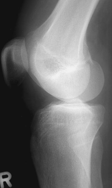
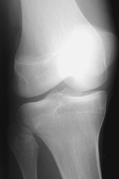
View knee radiographs: AP, Lateral, Oblique
View AP view.
 View lateral view.
 View oblique view.
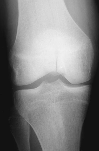
 AP, lateral, and oblique views of the knee are
obtained. There is a non-displaced intercondylar
fracture of the distal femur extending vertically. The
fracture is only appreciated on the AP view. These
radiographs demonstrate that it may be very difficult to
see some fractures at the wrong angle. If a fracture is
suspected, but not demonstrated on radiographs,
consider obtaining other views to more definitively
identify it.
An orthopedic surgeon was consulted by phone. He
was placed in a long leg splint and orthopedic follow-up
the next day was arranged.
Discussion
Fractures of the knee may be very obvious
clinically, but some of them are not. Most radiographs
of the knee will be normal, but it may be difficult to
identify small fractures of the knee with only two views.
Fractures of the distal femur are uncommon injuries.
These can be classified as supracondylar, condylar,
intercondylar and physeal. Most of these fractures are
large and are easily visible on AP and lateral
radiographs. In our patient's case, the intercondylar
fracture is small. Such intercondylar fractures often
extend further superiorly forming a "T" or "Y" shape as
they extend into the metaphysis of the distal femur.
Distal femur fractures are usually due to fairly
severe trauma. They may be associated with ipsilateral
hip fracture or dislocation, vascular injury, peroneal
nerve injury or damage to the quadriceps insertions.
References:
The Distal Femur (Chapter 17). In: Simon RR,
Koenigsknecht SJ. Emergency Orthopedics: The
Extremities, third edition. 1995, Norwalk, CT,
Appleton & Lange, pp. 267-272.
AP, lateral, and oblique views of the knee are
obtained. There is a non-displaced intercondylar
fracture of the distal femur extending vertically. The
fracture is only appreciated on the AP view. These
radiographs demonstrate that it may be very difficult to
see some fractures at the wrong angle. If a fracture is
suspected, but not demonstrated on radiographs,
consider obtaining other views to more definitively
identify it.
An orthopedic surgeon was consulted by phone. He
was placed in a long leg splint and orthopedic follow-up
the next day was arranged.
Discussion
Fractures of the knee may be very obvious
clinically, but some of them are not. Most radiographs
of the knee will be normal, but it may be difficult to
identify small fractures of the knee with only two views.
Fractures of the distal femur are uncommon injuries.
These can be classified as supracondylar, condylar,
intercondylar and physeal. Most of these fractures are
large and are easily visible on AP and lateral
radiographs. In our patient's case, the intercondylar
fracture is small. Such intercondylar fractures often
extend further superiorly forming a "T" or "Y" shape as
they extend into the metaphysis of the distal femur.
Distal femur fractures are usually due to fairly
severe trauma. They may be associated with ipsilateral
hip fracture or dislocation, vascular injury, peroneal
nerve injury or damage to the quadriceps insertions.
References:
The Distal Femur (Chapter 17). In: Simon RR,
Koenigsknecht SJ. Emergency Orthopedics: The
Extremities, third edition. 1995, Norwalk, CT,
Appleton & Lange, pp. 267-272.
Return to Radiology Cases In Ped Emerg Med Case Selection Page
Return to Univ. Hawaii Dept. Pediatrics Home Page