Hip and Knee Pain in a 4 Year Old
Radiology Cases in Pediatric Emergency Medicine
Volume 6, Case 15
Soledad S. U. Raroque, MD
Children's Medical Center of Dallas
University of Texas Southwestern School of Medicine
A four year old female is brought to the emergency
department with a complaint of right hip and knee pain.
About two hours prior to presentation, the child had
been running when she slipped and refused to bear
weight or move her right lower extremity. There is no
other history of significant medical or surgical problems.
Exam: VS: T 36.7, P120, RR 24, BP 120/77. She
is awake and alert in no acute distress. Her right lower
extremity is held in flexion at the hip and knee,
adducted and internally rotated. A bony prominence at
her right gluteal region is appreciated. She resists
attempts at passive range of motion about the hip
because of pain. Pulses are full. Motor and sensory
functions are all intact. The rest of her physical
examination is normal. Radiographs of the pelvis and
hips are obtained.
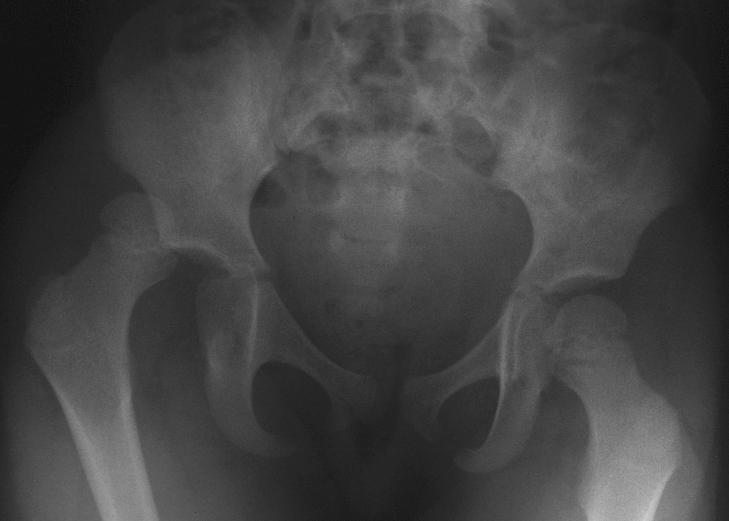
View Pelvis and Hip Radiographs.
AP view.
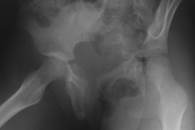
 Oblique view.
 The AP view shows a dislocation of the right hip.
The oblique view is difficult to interpret. Clinically, this
is a posterior dislocation. There is no evidence of
fracture. The patient was sedated in the emergency
department. Closed reduction of the right hip was done
by applying traction in line with the deformity and gently
flexing the hip to 90 degrees. Concentric reduction was
obtained. Repeat radiographs showed complete
reduction with no evidence of fracture or epiphyseal
injury.
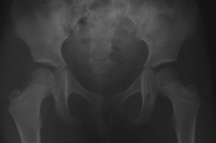
View Post-Reduction Pelvis and Hips.
AP view.
The AP view shows a dislocation of the right hip.
The oblique view is difficult to interpret. Clinically, this
is a posterior dislocation. There is no evidence of
fracture. The patient was sedated in the emergency
department. Closed reduction of the right hip was done
by applying traction in line with the deformity and gently
flexing the hip to 90 degrees. Concentric reduction was
obtained. Repeat radiographs showed complete
reduction with no evidence of fracture or epiphyseal
injury.
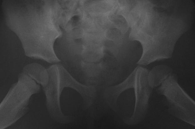
View Post-Reduction Pelvis and Hips.
AP view.
 Frog view.
 The child was hospitalized and underwent 48 hours
of skin traction. Upon discharge, she had continued full
range of motion, no pain and no evidence of
redislocation. She was followed-up by the orthopedic
service for several months.
Teaching Points:
1. Traumatic hip dislocation (THD) is an uncommon
injury in children and adolescents. Falls are the single
most common cause followed by high velocity injuries
such as motor vehicle accidents and sports-related
incidents. Boys are more frequently affected than girls.
It may also be seen in relatively minor trauma in young
children less than 5 years of age due perhaps to joint
laxity and a shallow acetabular fossa.
2. Posterior dislocations are more common than
anterior dislocations. These injuries often occur after a
blow to the knee with the hip and knee in flexion, as in
a motor vehicle crash with an unrestrained child striking
one knee against the dashboard. The leg would be
adducted, flexed and internally rotated at the hip, as in
this case. There is a relative shortening of the
extremity and protrusion of the greater trochanter into
the gluteal region. Anterior dislocations, on the other
hand, are usually caused by an excessive external
rotation or a direct blow to the greater trochanter with
the hip externally rotated. The leg would typically be
held in abduction, extension and external rotation.
3. The occurrence of an obvious traumatic episode
followed by limb dysfunction and local evidence of injury
of the affected body part narrows the differential
considerably. A plain radiograph may confirm a
fracture, avulsion, dislocation or soft-tissue injury. In
cases of THD, other views of the hip joint (e.g. oblique
and lateral views) may be difficult to obtain because of
the limited range of motion in some patients. Additional
radiographs of the ipsilateral extremity may be indicated
to rule out other fractures or injuries. Approximately
25% of hip dislocations are associated with knee
injuries.
4. Early recognition is essential in the management
of THD. Immediate closed reduction (within 6 hours
after the injury) under general anesthesia or
intravenous sedation has been shown to have a better
prognosis. A hip dislocation is reduced by flexing the
hip and knee to 90 degrees and applying axial traction
of the thigh. Repeat pelvic radiographs which show a
widened medial joint space indicate an incomplete
reduction. Late diagnosis and failure to achieve a
concentric reduction invariably requires an open
reduction. After a successful reduction, immobilization
either with traction or spica cast, may be done to
maintain stability. There is no consensus in the
orthopedic literature about the type of post-reduction
care or the duration of non-weight bearing significantly
affecting the prognosis of THD.
5. Computed tomography (CT) has been used after
reduction attempts to identify fractures or intraarticular
loose bodies not apparent on standard radiographs.
Magnetic resonance imaging (MRI) has also been
found to improve the diagnostic accuracy of hip
dislocation. Studies to compare the two modalities
have yet to be done.
6. The major complications of THD are frequent and
include avascular necrosis, recurrent dislocation, sciatic
nerve injury or traction injuries and traumatic arthritis.
The severity of the trauma and the period until
reduction are considered to be important prognostic
factors.
References
1. Attia MW, Gould JH. Traumatic hip dislocation in
a young child: A case report and discussion. Pediatric
Emergency Care. 1995;11(5):291-293.
2. Bachman D, Santora S. Orthopedic Trauma. In:
Fleisher GR, Ludwig S (eds). Textbook of Pediatric
Emergency Medicine, 3rd edition. Baltimore, MD,
Williams & Wilkins, 1993, pp. 1267-1268.
3. Huo MH, Root L, Buly RL, Mauri TM. Traumatic
Fracture-Dislocation of the Hip in a 2-Year-Old Child.
Orthopedics. 1992:15:1430-1433.
4. Moseley CF. Fractures and Dislocations of the
Hip. Instructional Course Lectures. 1992;41:397-401.
5. Poggi JJ, Callaghan JJ, Spritzer CE, et al.
Changes on Magnetic Resonance Images After
Traumatic Hip Dislocation. Clinical Orthopaedics and
Related Research. 1995;319:249-259.
6. Simon RR, Koenigsknecht SJ. The Hip, Pelvis
and Thighs. In: Emergency Orthopedics, 3rd edition.
Norwalk, CT, Appleton & Lange, 1995, pp. 430-435.
The child was hospitalized and underwent 48 hours
of skin traction. Upon discharge, she had continued full
range of motion, no pain and no evidence of
redislocation. She was followed-up by the orthopedic
service for several months.
Teaching Points:
1. Traumatic hip dislocation (THD) is an uncommon
injury in children and adolescents. Falls are the single
most common cause followed by high velocity injuries
such as motor vehicle accidents and sports-related
incidents. Boys are more frequently affected than girls.
It may also be seen in relatively minor trauma in young
children less than 5 years of age due perhaps to joint
laxity and a shallow acetabular fossa.
2. Posterior dislocations are more common than
anterior dislocations. These injuries often occur after a
blow to the knee with the hip and knee in flexion, as in
a motor vehicle crash with an unrestrained child striking
one knee against the dashboard. The leg would be
adducted, flexed and internally rotated at the hip, as in
this case. There is a relative shortening of the
extremity and protrusion of the greater trochanter into
the gluteal region. Anterior dislocations, on the other
hand, are usually caused by an excessive external
rotation or a direct blow to the greater trochanter with
the hip externally rotated. The leg would typically be
held in abduction, extension and external rotation.
3. The occurrence of an obvious traumatic episode
followed by limb dysfunction and local evidence of injury
of the affected body part narrows the differential
considerably. A plain radiograph may confirm a
fracture, avulsion, dislocation or soft-tissue injury. In
cases of THD, other views of the hip joint (e.g. oblique
and lateral views) may be difficult to obtain because of
the limited range of motion in some patients. Additional
radiographs of the ipsilateral extremity may be indicated
to rule out other fractures or injuries. Approximately
25% of hip dislocations are associated with knee
injuries.
4. Early recognition is essential in the management
of THD. Immediate closed reduction (within 6 hours
after the injury) under general anesthesia or
intravenous sedation has been shown to have a better
prognosis. A hip dislocation is reduced by flexing the
hip and knee to 90 degrees and applying axial traction
of the thigh. Repeat pelvic radiographs which show a
widened medial joint space indicate an incomplete
reduction. Late diagnosis and failure to achieve a
concentric reduction invariably requires an open
reduction. After a successful reduction, immobilization
either with traction or spica cast, may be done to
maintain stability. There is no consensus in the
orthopedic literature about the type of post-reduction
care or the duration of non-weight bearing significantly
affecting the prognosis of THD.
5. Computed tomography (CT) has been used after
reduction attempts to identify fractures or intraarticular
loose bodies not apparent on standard radiographs.
Magnetic resonance imaging (MRI) has also been
found to improve the diagnostic accuracy of hip
dislocation. Studies to compare the two modalities
have yet to be done.
6. The major complications of THD are frequent and
include avascular necrosis, recurrent dislocation, sciatic
nerve injury or traction injuries and traumatic arthritis.
The severity of the trauma and the period until
reduction are considered to be important prognostic
factors.
References
1. Attia MW, Gould JH. Traumatic hip dislocation in
a young child: A case report and discussion. Pediatric
Emergency Care. 1995;11(5):291-293.
2. Bachman D, Santora S. Orthopedic Trauma. In:
Fleisher GR, Ludwig S (eds). Textbook of Pediatric
Emergency Medicine, 3rd edition. Baltimore, MD,
Williams & Wilkins, 1993, pp. 1267-1268.
3. Huo MH, Root L, Buly RL, Mauri TM. Traumatic
Fracture-Dislocation of the Hip in a 2-Year-Old Child.
Orthopedics. 1992:15:1430-1433.
4. Moseley CF. Fractures and Dislocations of the
Hip. Instructional Course Lectures. 1992;41:397-401.
5. Poggi JJ, Callaghan JJ, Spritzer CE, et al.
Changes on Magnetic Resonance Images After
Traumatic Hip Dislocation. Clinical Orthopaedics and
Related Research. 1995;319:249-259.
6. Simon RR, Koenigsknecht SJ. The Hip, Pelvis
and Thighs. In: Emergency Orthopedics, 3rd edition.
Norwalk, CT, Appleton & Lange, 1995, pp. 430-435.
Return to Radiology Cases In Ped Emerg Med Case Selection Page
Return to Univ. Hawaii Dept. Pediatrics Home Page