Smartphones can be used to estimate body composition and indicate health and mortality risks, according to a new study by a University of Hawaiʻi Cancer Center researcher and his team. This accessible and cost-effective option will enable early screening and monitoring of physiological indicators of metabolic diseases in regions where medical imagery or clinical assessment is not available.
“This is beneficial as smartphones are already owned by middle and low-income individuals who are most susceptible to metabolic diseases, including diabetes and non-alcoholic fatty liver disease,” said John Shepherd, lead study investigator.
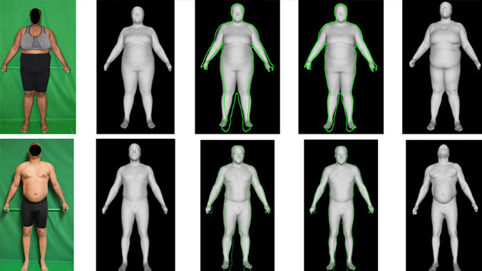
Patients’ health and mortality risks can be indicated by total and regional body composition measurements using 3D imaging, which require expensive and specialized equipment and are restricted to clinical settings. The study found that body composition can be estimated from a single frontal consumer level photograph of a patient’s body that can be taken on conventional consumer cameras, such as those on smartphones.
Photographs taken of patients are processed to predict internal health measures such as body fat percentage and lean muscle mass, which provides useful and detailed information about various health and wellness risks.
This new technology helps to achieve the UH Cancer Center’s mission of reducing the burden of cancer through research, education, patient care and community outreach because of its solution to high-cost medical treatment that is not feasible for many of Hawaiʻi’s patients in lower socioeconomic brackets.