Using an innovative new approach to sampling corals, researchers at the University of Hawaiʻi at Mānoa are now able to create maps of coral biochemistry that reveal with unprecedented detail the distribution of compounds that are integral to the healthy functioning of reefs. The study was published in Communications Biology.
“This work is a major step in understanding the coral holobiont [the coral animal and all of its associated microorganisms], which is critical for reef restoration and management,” said lead author Ty Roach, who conducted this study as a postdoctoral researcher at the Hawaiʻi Institute of Marine Biology (HIMB) in the UH Mānoa School of Ocean and Earth Science and Technology.
Despite occupying a tiny fraction of the ocean, coral reefs are one of the most diverse and productive ecosystems on the planet and provide critical habitats for many species and protection for coastal communities.
Biochemicals, such as amino acids, compounds that affect development and growth, and others that have antibacterial or antioxidant properties, have a direct relation to how resilient coral will be in the face of stressors, such as warmer ocean temperatures and ocean acidification.
Evaluating individual polyps
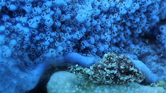
The team of HIMB researchers developed a method to investigate a single coral polyp at a time.
“This new technique allows us to sample corals in a way that is much less invasive and damaging than the previous methods, meaning that we can now take more samples and repeat sampling efforts more often with less damage to the coral,” Roach added.
Using sophisticated chemical analyses, the team determined the exact biochemicals that are contained in each individual polyp and mapped them back to their respective location in the coral colony. With this, they created maps of biochemicals in corals across multiple spatial scales—from individual, 1 millimeter-wide polyps to 100 meter-long reefs.
“The use of this technique across these scales allowed us to discover a strong biochemical signature that identifies polyps as being from a single colony, a weaker signature between branches within colonies, and variation along the branches that is related to where the polyp came from on the branch,” said Roach. “Surprisingly, this method was even able to discriminate between adjacent polyps with very high rates of accuracy.”
Mapping biochemicals
By mapping biochemicals back to their source—from either the coral animal or its symbiotic algae—the researchers also determined that the compounds in the polyps were mainly driven by molecules that came from the coral instead of the algae.
“Importantly, this work tells us how corals structure their biochemicals across different scales of interest, which is critical for design, analysis and interpretation of studies on coral reef biochemistry,” added Roach. “We plan to use this approach in future studies to map the temporal and spatial distribution of coral biomolecules in ways that were not previously possible without damaging whole coral colonies.”
–By Marcie Grabowski